Skip to main navigationSkip to main content
The University of Southampton
Engineering

Research project: A novel way to enhance the diagnosis of early stage of colorectal cancer through micro-computed tomography imaging

Currently Active: 
Yes

Colorectal cancer (CRC) is currently the second most common cause of cancer-related death in Europe. Early diagnosis of CRC is essential as it leads to better therapy and survival rates. However, advanced cancers are still being reported, although patients are screened by regular surveillance colonoscopies and biopsies. In recent years, advanced technologies such as gene microarrays have attempted to find susceptible genes to link the inammation and CRC for diagnostic and prediction purposes, but this has not been very successful. Patients with long-term inammatory bowel disease (IBD) in particular, have a 20% lifetime risk of developing CRC. To this end, a novel way is proposed, to enhance the timely diagnosis of early-stage CRC in patients with IBD or familial heritage through micro-computed tomography.
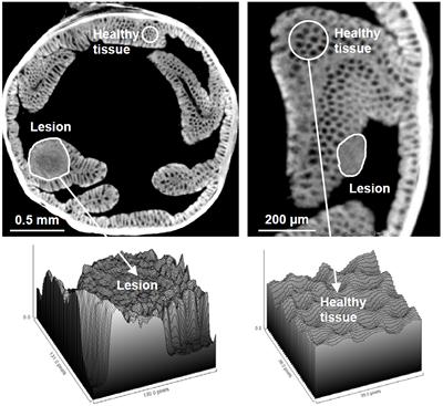
Diagram
Figure 1

During previous work in the group, a new sample preparation protocol for colon tissue based on phosphotungstic acid as X-Ray contrast agent, was developed and optimised for microCT imaging. Following this novel approach, excised colon tissue was imaged in 3D by high-resolution synchrotron-based CT at the TOMCAT beamline of the Swiss Light Source and using a state-of-the-art lab microCT system (Versa 510, Xradia/Zeiss) at the X-Ray Imaging Centre muVIS of the University of Southampton, at spatial resolutions between 1-5 um. Lesions (loss of crypts and goblet cells at the mucosa layer) could be detected and visualised during early-stage CRC (see Figure 1), which resembled aberrant crypt foci that are reported to be related to tumorigenesis. First comparisons with histological results suggest that it is possible to identify aberrant crypt foci based on height profiles as pre-dysplastic lesions that can be seen in early-stage CRC.

In this PhD project, a new microCT protocol for imaging micro-structures of un-contrasted colon tissue will be developed. By the end of the project, it should be possible that standard histopathological wax blocks are scanned indestructively at some point within the standard histopathological workflow.

Additional diagnostic information such as 3D shapes, surface textures, etc., which are elements only visible in the 3D internal structure of the tissue will then be available. Supplying all this information to histopathologists as an extra assistive new tool, will provide a whole new insight in the diagnosis of early stage colorectal cancer.

Related research groups

Bioengineering Science
Share this research project Share this on Facebook Share this on Twitter Share this on Weibo
Privacy Settings